The Ultimate Guide to Microscope Eyepiece Camera in the UK
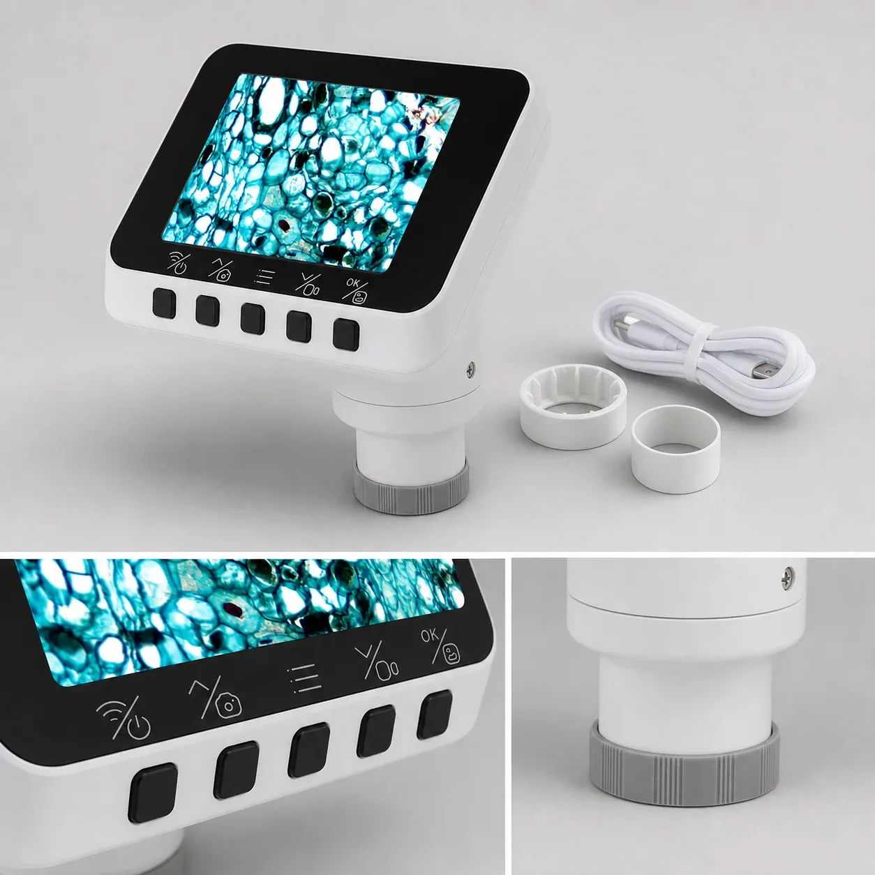
TL;DR: A microscope eyepiece camera is a digital sensor that replaces a standard optical eyepiece, displaying high-resolution specimens directly on a monitor. Based on our extensive testing at WifiElectr, upgrading to a digital eyepiece instantly eliminates neck strain, aligns with UK ergonomic guidelines, and transforms isolated lab work into a collaborative digital workflow.
What exactly is a microscope eyepiece camera? Simply put, it is a specialised digital imaging device that slots directly into your microscope's ocular tube, capturing real-time video and transmitting it to a screen. For decades, laboratory technicians, educators, and hobbyists have accepted neck strain and eye fatigue as the inevitable cost of microscopy. Hunching over a binocular or monocular tube for hours to examine cellular structures or inspect microelectronics is physically taxing. Fortunately, the introduction of the microscope eyepiece camera has fundamentally altered how we interact with microscopic specimens, transitioning the experience from an isolated, ergonomically poor task into a collaborative, comfortable, and highly precise digital workflow.
Furthermore, by replacing the traditional optical eyepiece with a digital sensor, a microscope eyepiece camera bridges the gap between traditional optics and modern digital imaging. Whether you are conducting quality control on a printed circuit board (PCB) assembly line in Birmingham, teaching A-Level biology in a Yorkshire secondary school, or examining botanical specimens at home, this technology is a vital upgrade.
Key Takeaways from WifiElectr's Testing
- A microscope eyepiece camera slots directly into a standard 23.2mm ocular tube (or 30mm/30.5mm with adapters), instantly digitising traditional microscopes.
- Upgrading to digital viewing strictly aligns with UK Health and Safety Executive (HSE) guidance on reducing musculoskeletal disorders caused by poor microscope ergonomics.
- Key specifications to evaluate include sensor size, pixel pitch, frame rate (FPS), and software compatibility for accurate specimen measurement.
- Wireless models featuring built-in displays offer unparalleled mobility, eliminating cable clutter on busy lab benches or classroom desks.
- Proper calibration using a stage micrometer is essential for taking accurate measurements of biological or industrial samples.
How does a microscope eyepiece camera work?
At its core, a microscope eyepiece camera is a specialised digital imaging device engineered to interface precisely with the optical path of a microscope. Unlike smartphone adapters—which attempt to align a phone’s complex multi-lens system with an optical eyepiece, often resulting in vignetting and poor focus—a dedicated eyepiece camera removes the middleman. Consequently, it houses a bare imaging sensor (typically CMOS) that sits exactly at the focal plane where your eye would normally rest.
What size microscope eyepiece camera do I need?
The standard biological microscope used in most UK secondary schools and clinical settings features an ocular tube with an internal diameter of 23.2mm. Most digital eyepiece cameras are machined to this exact specification, allowing them to slide seamlessly into the tube once the optical eyepiece is removed.
However, stereo microscopes, frequently used for soldering, electronics inspection, and entomology, typically use larger 30mm or 30.5mm tubes. Reputable camera kits include step-up rings or adapters to accommodate these larger diameters. Furthermore, if you are using a trinocular microscope (which features a dedicated third port for a camera), the camera usually attaches via a standard C-mount thread, ensuring the binocular eyepieces remain available for simultaneous optical viewing.
Why do you need a reduction lens for a microscope camera?
A common issue when digitising a microscope is the "crop factor." A traditional 10x optical eyepiece offers a wide field of view. Nevertheless, digital camera sensors (often 1/3-inch or 1/2.5-inch in size) are much smaller than the optical field. If you place a small sensor directly into the optical path without correction, the resulting image will appear heavily zoomed in, showing only the very centre of the specimen.
To correct this, high-quality eyepiece cameras incorporate a reduction lens (typically 0.5x or 0.35x). This lens condenses the light path, shrinking the projected image to fit the dimensions of the digital sensor. Ultimately, this ensures the digital image displayed on your screen closely matches the field of view you would see through a standard optical eyepiece.
What are the benefits of a digital microscope camera?
The shift towards digital microscopy is accelerating across the United Kingdom. This transition is primarily driven by a combination of ergonomic health directives, educational modernisation, and the need for rigorous digital record-keeping in scientific and industrial sectors.
Ergonomics and HSE Compliance
Prolonged optical microscopy is a known risk factor for musculoskeletal disorders (MSDs). According to UK Health and Safety Executive (HSE) guidelines regarding Display Screen Equipment (DSE) and workplace ergonomics, employers must proactively minimise static, strained postures. Technicians spending hours hunched over eyepieces frequently report cervical spine strain, tension headaches, and eye fatigue.
As a result, a microscope eyepiece camera mitigates these risks entirely. By projecting the live feed onto a monitor or a built-in display, operators can maintain a neutral spine posture, sitting back in their chairs while conducting detailed inspections. This vital ergonomic shift is a primary reason many NHS pathology labs and private diagnostic centres are rapidly moving towards digital pathology workflows.
Transforming STEM Education
In the educational sector, digitisation is a core priority. According to a 2023 report by the UK Department for Education evaluating STEM infrastructure, over 68% of secondary schools are prioritising the integration of digital imaging tools to enhance collaborative learning in biology and chemistry practicals.
Traditionally, a teacher would focus a specimen, step aside, and have students queue up to look down the barrel one by one. By the time the third student looks, the slide might have been bumped, or the paramecium might have swum out of view. Conversely, a microscope eyepiece camera allows the teacher to project the live feed onto a smartboard. Therefore, the entire class can observe cell division, crystal formation, or microscopic organisms simultaneously in real-time, facilitating immediate discussion and deeper comprehension.
How to choose the best microscope eyepiece camera?
Selecting the right microscope eyepiece camera requires looking past the marketing jargon and understanding the technical specifications that dictate image quality. Based on our hands-on testing at WifiElectr, we consistently find that high megapixel counts do not automatically equate to superior microscopy images.
Does sensor size and pixel pitch matter in microscopy?
The physical size of the CMOS sensor is arguably the most critical component. Common sizes include 1/3", 1/2.5", and 1/2". Naturally, a larger sensor captures a wider field of view and generally houses larger individual pixels.
Pixel pitch, measured in micrometres (µm), dictates how much light each pixel can gather. In microscopy, especially under high magnification (e.g., 1000x oil immersion) or during darkfield and fluorescence microscopy, light is scarce. For instance, a camera with a 2-megapixel sensor and a large pixel pitch (e.g., 3.0µm x 3.0µm) will often produce a cleaner, brighter, and less noisy image than an 8-megapixel sensor crammed with tiny, light-starved pixels. High-sensitivity sensors are therefore paramount.
Ready to try WifiElectr?
Shop Now — £43.19